


Files
Download Full Text (4.5 MB)
Abstract
Motor Evoked Potential Recruitment Curves Indicate Neuroplasticity after Spinal Cord Injury
Yasmina Zeineddine, Depts. of Biomedical Engineering and Anthropology, with Thibault Roumengous, Graduate Student in Biomedical Engineering, and Dr. Carrie Peterson, Dept. of Biomedical Engineering
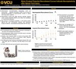
Introduction: Motor evoked potential (MEP) recruitment curves in response to transcranial magnetic stimulation across a range of stimulation intensities can provide insight into the condition of neural pathways to a muscle. Further, corticomotor reorganization associated with recovery of motor function may be reflected in changes in the MEP recruitment curve. At low TMS intensity, the MEP often consists of a single direct wave, whereas at higher stimulus intensities, the MEP amplitude increases due to recruitment of later indirect waves (I-waves). These late I-waves are thought to depend on trans-synaptic activation of corticospinal axons through excitatory interneurons. [1] In impaired patients, MEP recruitment curves can inform the development of individualized rehabilitation treatments, as curve irregularities can reveal specific deficits, and enhance our understanding of the neuroplastic changes that occur after injury. In comparing the biceps brachii recruitment curve of subjects with cervical spinal cord injury (SCI) to nonimpaired individuals, we hypothesized that individuals with SCI would have greater biceps excitability, and therefore greater RC slopes, due to the biceps cortical representation growing in response to triceps paralysis [2]. Materials and Methods: Ten nonimpaired (4 female, 6 male) and 5 impaired (3 female, 2 male) subjects consented to participate in the study. Subjects were seated and had their dominant arm positioned at a 90° elbow angle. EMG surface electrodes were placed on the biceps after being cleaned with alcohol wipes. The biceps cortical hotspot was determined as the area over the motor cortex where TMS evoked the largest MEP response. The resting motor threshold (RMT) was the lowest stimulus intensity required to elicit a 50μVpp MEP response in 3/5 trials. TMS was performed with a 126 mm diameter double cone coil and Magstim BiStim2 . MEPs were recorded from the BB and normalized by dividing by Mmax. Recruitment curves were recorded at stimulus intensities ranging from 80%- 160% of subject RMT in 10% increments. Pulse intensities were randomly administered with interstimulus intervals of 10 s. The data was recorded using Spike software and processed in Matlab. MEPs were excluded if they exceeded ±3 standard deviations of the mean response per intensity. Results and Discussion: The recruitment curve slopes for individuals with SCI, on average, were greater relative to the slopes of nonimpaired individuals. This was based on an analysis of MEPs between intensities of 100% and 140% RMT, wherein the slope was on average 5.13 across individuals with SCI, and 1.49 in the nonimpaired population. The greater slope in individuals with SCI suggests enhanced excitability of the biceps, which is consistent with previous studies showing greater cortical representation of non-paralyzed hand muscles relative to paralyzed muscles [2]. Conclusions: Our results indicate that cervical SCI promotes greater excitability in the muscles controlled by nerves rostral to the location of injury, and demonstrate neural plasticity following injury. The steeper slopes in individuals with SCI indicate greater recruitment of later I-waves. Whether increased recruitment of later I-waves is associated with greater cortical map area is unclear and will require further investigation.
Publication Date
2020
Faculty Advisor/Mentor
Carrie Peterson, Ph.D.
Sponsorship
Virginia Commonwealth University. Undergraduate Research Opportunities Program
Is Part Of
VCU Undergraduate Research Posters
Rights
© The Author(s)