


Preview
Date
2001
Type of Abnormality
Radiographic abnormality
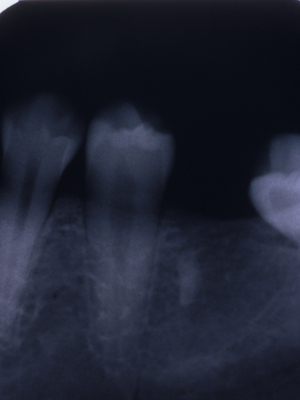
Location of Abnormality
Mandibular alveolar ridge distal to the root of the second premolar
Description
Radiopaque area of the alveolar ridge with the structural appearance of the apical portion of a tooth root. A thin periodontal ligament space is visible on the distal. Note the similarity of the radiodensity of the root tip and the adjacent premolar root
Topical Subject
Jaws -- Diseases
Medical Subject
Pathology, Oral; Tooth Root -- radiography; Mandible -- radiography; Mandible -- pathology
Original Item Medium
35 mm slide
Genre
slides (photographs)
Local Genre
photograph
Type
Still Image
Digital Format
image/jpg
Language
eng
Rights Statement URL
Rights
This material is protected by copyright, and the copyright is held by VCU. You are permitted to use this material in any way that is permitted by copyright. In addition, this material is licensed under a Creative Commons Attribution-Noncommercial-Share Alike 4.0 International license (CC BY-NC-SA 4.0). Acknowledgment of Virginia Commonwealth University Libraries as a source is required.
Collection
Oral Pathology Review Images
Contributors
Page, Dennis; Virginia Commonwealth University. Dept. of Oral Pathology; Virginia Commonwealth University. School of Dentistry. Dept. of Oral Pathology; Medical College of Virginia. School of Dentistry
Source
VCU School of Dentistry Oral Pathology Slides, RG 4-5: University Archives Vice President for Health Sciences, Dean, School of Dentistry, Special Collections and Archives, VCU Health Sciences Library, Virginia Commonwealth University, Richmond, Va.
File Name
opr53.jpg