


Files
Download Full Text (517 KB)
Abstract
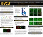
The response of endothelial cells, innermost layer of blood vessels, to blood flow is thought to be critical in the initiation and progression of atherosclerosis. Atherosclerosis in the human body is non-random and is highly correlated to vessel sites which experience oscillatory and reversing blood flow. Endothelial cells (ECs), the inner most cell layer of blood vessels are highly responsive to the drag force from blood flow, known as shear stress. To study endothelial cell responses to shear stress we used a parallel plate flow chamber in which we exposed endothelial cells to defined fluid shear stress. Using fluorescence resonance energy transfer (FRET)-based biosensors we examined both how zinc levels change in response to fluid shear stress, as well as the mechanical forces applied onto the nucleus. Previous work by the lab suggested that zinc levels were increased following exposure to 24 hours of fluid shear stress. To better understand these changes we utilized four zinc-FRET sensors specific to zinc levels in the nucleus, Golgi, endoplasmic reticulum, and cytoplasm. Although our control experiments indicated the sensors were working properly, we did not observe consistent changes in zinc levels in any of these four subcellular locations for ECs exposed to 24 hours fluid shear stress as compared to static controls. Next we investigated how fluid shear stress affects forces applied to the nucleus. Our laboratory recently developed a nesprin-2 force sensor that has provided the first direct evidence that the nucleus of a cell is subject to mechanical force. Additionally, the rare advanced aging disease Hutchinson-Gilford Progeria syndrome (HGPS), is associated with rapid onset of atherosclerosis despite the absence of standard risk factors (obesity, high cholesterol, etc). HGPS results from a mutation in lamin A that creates an altered nuclear shape. Therefore we hypothesize that forces on the nucleus are a critical mediator of the EC response to shear stress, and that HGPS cells may have altered mechanotransduction that promotes atherosclerosis. We have shown that HGPS cells grown in static culture have reduced mechanical force on the nucleus. Additionally we have shown that ECs expressing the HGPS mutant lamin A do not respond properly to shear stress. We are currently using the nesprin-2 tension sensor to measure mechanical forces on ECs subjected to shear stress.
Publication Date
2015
Subject Major(s)
Biomedical Engineering
Disciplines
Molecular, Cellular, and Tissue Engineering
Current Academic Year
Junior
Faculty Advisor/Mentor
Dr. Daniel Conway
Rights
© The Author(s)