


Files
Download Full Text (2.9 MB)
Abstract
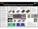
Finite Element Analysis of 3D-printed PCL Scaffolds for Synergizing Cellular Micro-Environment and Mechanical Stimuli to Enhance Engineered Tissue Growth in Vitro
Ireolu Orenuga,1 Phillip Glass,2 Daeha Joung,2 Joao S. Soares1
- Department of Mechanical and Nuclear Engineering, College of Engineering, Virginia Commonwealth University
- Department of Physics, College of Humanities & Sciences, Virginia Commonwealth University
Introduction: Tissue engineering aims to create viable and functional engineered tissues via biodegradable scaffolds and autologous cells. Scaffolds play an essential part in organizing the architecture of developing tissues and aid in the proper function of implants acutely by serving as mechanical support and long-term by degrading and undergoing absorption as de novo tissue is produced. Polycaprolactone (PCL) is a commonly used biodegradable and biocompatible material. PCL scaffolds are typically electrospun or nonwoven, which produces random microstructures without a very robust control. 3D printed PCL scaffolds allow for the design of a structured and controlled cellular micro-environment and mechanical properties for cells to grow in vitro. However, the translation of mechanical training at the tissue level happening in bioreactors during in vitro culture to cellular stimulation is poorly understood. Finite Element Analysis (FEA) of 3d-printed PCL scaffolds may elucidate the different roles of microstructural parameters in cellular mechanotransduction and provide insight on how to effectively engineer better tissues.
Methods: SolidWorks was used to make four models of 3D-printed PCL fibers with 0-90 and 45-45 orientations in two configurations, i.e. aligned and staggered fibers. The completed 0-90 and 45-45 orientations staggered model was exported as an .stl file to 3D-print. SEM imaging analyzing the 3d-printing results were conducted to obtain realistic microstructures of the 3D-printed PCL scaffolds. These 3D-models were imported into FEBio for FEA analysis of the deformations involved with in vitro mechanical training up to a uniaxial strain of 50% (aligned with the 0-degree orientation). Additionally, we have performed exploratory cell seeding experiments on some scaffolds to analyze seeding efficiency and mechanical testing of virgin scaffolds to validate the FEA results.
Results: The 3D-printed models produced were a 0-90 and a 45-45 staggered configuration scaffold each with dimensions of 8 x 20 x 0.45mm thickness with 16 layers. PCL fibers were printed as ribbons with 10 by 85 microns and spaced apart creating pores of 235 by 235 microns. Meshing both configurations was successful and FEA simulations showed that scaffolds can undergo strains of up to 33% before pores start collapsing. Mechanical tests showed the scaffold was able to undergo strains of up to 10% in the elastic regime without appreciating damage. Drip seeding experiments showed that RVSMCs are able to survive and grow on the 3D printed PCL scaffolds.
Conclusions: FEA has proven to be useful for predicting mechanical behavior of 3D printed PCL scaffolds. Future experiments will focus on conducting mechanical testing on more scaffolds and in vitro engineered tissue culture under mechanical stimulation.
Publication Date
2023
Keywords
Tissue engineering, Biomedical, Biomaterials, Biocompatibility, 3D-Printing, Mechanical Structures, FEA
Disciplines
Biomaterials | Biomechanical Engineering | Biomedical Engineering and Bioengineering | Computer-Aided Engineering and Design | Mechanical Engineering | Molecular, Cellular, and Tissue Engineering
Current Academic Year
Senior
Faculty Advisor/Mentor
Joao Soares
Faculty Advisor/Mentor
Daeha Joung
Rights
© The Author(s)
Included in
Biomaterials Commons, Biomechanical Engineering Commons, Computer-Aided Engineering and Design Commons, Molecular, Cellular, and Tissue Engineering Commons